
Self-organized gold nanoparticles on SiO2 surface nanopatterns induced by MeV ion implantation
DOI:
https://doi.org/10.47566/2025_syv38_1-250101Keywords:
Ion implantation, Surface nanostructures, Gold nanoparticles, Surface plasmon resonanceAbstract
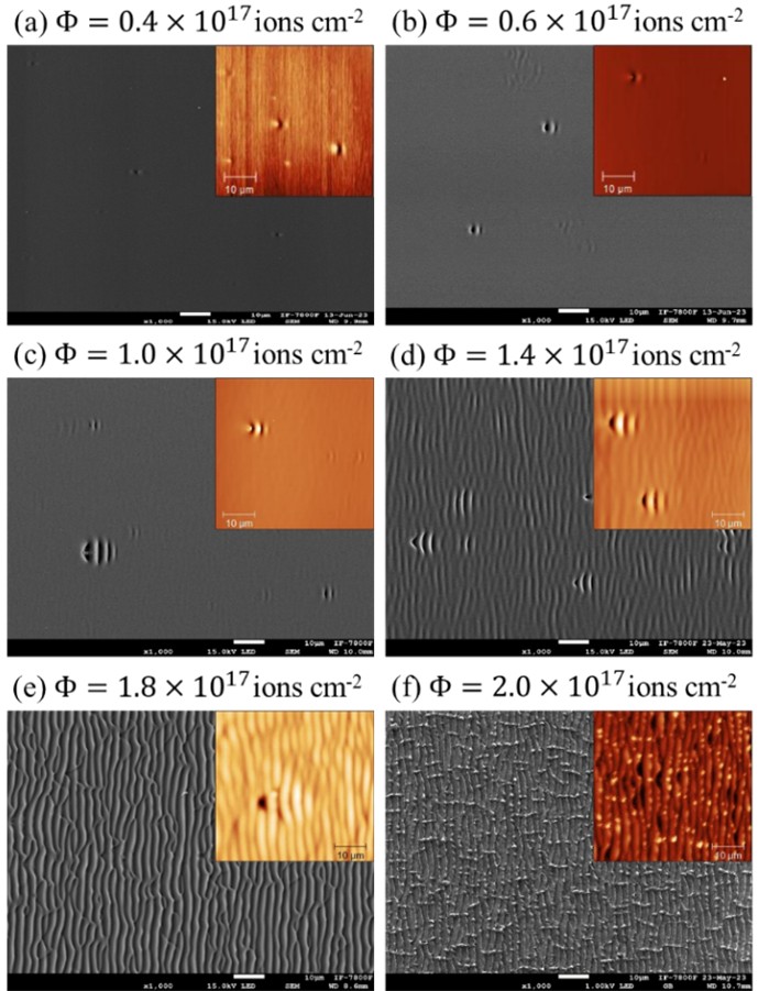
Ion implantation at MeV energies leads to the formation of surface nano-patterns on diverse kinds of materials, such as metals, dielectrics, and semiconductors. Furthermore, under oblique irradiation, the implanted ions are preferentially deposited on the crests of such patterns. Based on previous experimental results, in this work the formation of gold nanoparticles is produced and found to be organized onto prepatterned surfaces as produced by 1.8 MeV Au ion implantation on SiO2 substrates at an impinging angle of 45°. The ion implantation was performed with an ion beam current of 400 nA for a total ion fluence between 0.2 ×1017 cm-2 and 2.0 ×1017 cm-2. After implantation, to assist gold nanoparticle nucleation, substrates were annealed at 1000 °C. The surface morphology and topography of the implanted substrates were analyzed by atomic force microscopy (AFM) and scanning electron microscopy (SEM), respectively. For the lowest fluences (0.4 - 0.8 ×1017 cm-2), the formation of isolated surface structures on a flat background was observed whose morphology mimics the exoskeleton of a bug. According to SEM-EDS analysis, the formation of gold nanoparticles on the exoskeleton of the bugs was observed. For intermediate fluences (0.8 - 1.4 ×1017 cm-2), a ripple background now appears and begins to surround the bugs. At the highest fluences (1.4 - 2.0 ×1017 cm-2), the surface shows only a ripple pattern. In this last case, the gold nanoparticles are formed along the crests of the surface ripples. Finally, the optical absorption of the samples (except the one corresponding to the lowest fluence) shows the presence of a localized surface plasmon resonance (LSPR) in the wavelength region of 520 - 540 nm, suggesting the presence of gold nanoparticles.
Downloads
References
. C.F. Cruz-Garcia, J Rickards, M.A. Garcia, L.R. de la Vega, J Cañetas-Ortega, J.G. Morales-Morales, L. Rodríguez-Fernández, Phys. Scr. 98, 105956 (2023).
https://doi.org/10.1088/1402-4896/acf7fe
. M.A. Garcia, R. Martínez-Cervantes, J. Rickards, J. Cañetas-Ortega, J.G. Morales-Morales, L.R. de la Vega, L. Rodríguez-Fernández, Nucl. Instrum. Methods Phys. Res. B 550, 165304 (2024).
https://doi.org/10.1016/j.nimb.2024.165304
. U. Valbusa, C. Boragno, F. Buatier de Mongeot, J. Phys. Condens. Matter 14, 8153 (2002).
https://doi.org/10.1088/0953-8984/14/35/301
. J. Muñoz-García, L. Vázquez, M. Castro, R. Gago, A. Redondo-Cubero, A. Moreno-Barrado, R. Cuerno, Mater. Sci. Eng. R 86, 1 (2014).
https://doi.org/10.1016/j.mser.2014.09.001
. R. Cuerno, J.S. Kim, J. Appl. Phys. 128, 180902 (2020).
https://doi.org/10.1063/5.0021308
. C. Mennucci, S. Del Sorbo, S. Pirotta, M. Galli, L.C. Andreani, C. Martella, M.C. Giordano, F. Buatier de Mongeot, Nanotechnology 29, 355301 (2018).
https://doi.org/10.1088/1361-6528/aac9ac
. M.C. Giordano, F. Buatier de Mongeot, Adv. Mater. 30, 1801840 (2018).
https://doi.org/10.1002/adma.201801840
. W.L. Chan, E. Chason, J. Appl. Phys. 101, 121301 (2007).
https://doi.org/10.1063/1.2749198
. M.A. Garcia, R. Gago, J. Rickards, R. Trejo-Luna, J. Cañetas Ortega, L.R. de la Vega, L. Rodríguez-Fernández, J. Phys. Condens. Matter 30, 274005 (2018).
https://doi.org/10.1088/1361-648X/aac7f6
. S.K. Srivastava, K.G.M. Nair, R. Kamalakannan, M. Kamruddin, B.K. Panigrahi, A.K. Tyagi, AIP Conf. Proc. 1447, 741 (2012).
https://doi.org/10.1063/1.4710216
. R.M. Bradley, J.M.E. Harper, J. Vac. Sci. Technol. A 6, 2390 (1988).
https://doi.org/10.1116/1.575561
. B. Davidovitch, J. Aziz, P. Brenner, Phys. Rev. B 76, 205420 (2007).
https://doi.org/10.1103/PhysRevB.76.205420
. B. Davidovitch, J.M. Aziz, M.P. Brenner, J. Phys. Condens. Matter 21, 224019 (2009).
https://doi.org/10.1088/0953-8984/21/22/224019
. R.M. Bradley, H. Hofsäss, J. Appl. Phys. 120, 074302 (2016).
https://doi.org/10.1063/1.4960807
. B. Rauschenbach, Low-Energy Ion Irradiation of Materials: Fundamentals and Application, 1st ed. (Springer Cham, 2022) p. 250-251.
https://doi.org/10.1007/978-3-030-97277-6
. C.C. Umbach, R.L. Headrick, K.C. Chang, Phys. Rev. Lett. 87, 246104 (2001).
https://doi.org/10.1103/PhysRevLett.87.246104
. M. Castro, R. Cuerno, Appl. Surf. Sci. 258, 4171 (2012).
https://doi.org/10.1016/j.apsusc.2011.09.008
. H. Gnaser, Low Energy Ion Irradiation of Solid Surfaces, 1 st ed. (Springer-Verlag, 1999) p. 153-204.
https://doi.org/10.1007/BFb0110693
. L. Douillard, J.P. Duraud, Nucl. Instrum. Methods Phys. Res. B 107, 212-217 (1996).
https://doi.org/10.1016/0168-583X(95)01044-0
. W. Ou, B. Zhou, J. Shen, C. Zhao, Y.Y. Li, J. Lu, iScience 24, 101982 (2021).
https://doi.org/10.1016/j.isci.2020.101982
. P. Long Truong, X. Ma, S.J. Sim, Nanoscale 6, 2307 (2014).
https://doi.org/10.1039/C3NR05211G
. J. Liu, H. He, D. Xiao, S. Yin, W. Ji, S. Jiang, D. Luo, B. Wang, Y. Liu, Materials (Basel) 11, 1833 (2018).
http://doi.org/10.3390/ma11101833
. T.Y. Jeon, D.J. Kim, S.G. Park, S.H. Kim, D.H. Kim, Nano Convergence 3, 18 (2016).
https://doi.org/10.1186/s40580-016-0078-6
. J.P. Cordero-Santiago, A. Crespo-Sosa, Photonics Nanostructures: Fundam. Appl. 51, 101051 (2022).
https://doi.org/10.1016/j.photonics.2022.101051
. A. Chhatre, P. Solasa, S. Sakle, R. Thaokar, A. Mehra, Colloids Surf. A Physicochem. Eng. Asp. 404, 83 (2012).
https://doi.org/10.1016/j.colsurfa.2012.04.016
. X. Ou, Y. Liu, M. Zhang, L. Hua, S. Zhan, Mikrochim. Acta 188, 304 (2021).
https://doi.org/10.1007/s00604-021-04964-1
. H. Duan, T.Wang, Z. Su, H. Pang, C. Chen, Nanotechnol. Rev. 11, 1 (2022).
https://doi.org/10.1515/ntrev-2022-0039
. P. Mandal, S. Sharma, Renew. Sustain. Energy Rev. 65, 537 (2016).
https://doi.org/10.1016/j.rser.2016.07.031
. M. Saini, S. Augustine, M. Ranjan, T. Som, Appl. Surf. Sci. 512, 145703 (2020).
https://doi.org/10.1016/j.apsusc.2020.145703
. D. Ne?as, P. Klapetek, Central Eur. J. Phys. 10, 181 (2012).
https://doi.org/10.2478/s11534-011-0096-2
. M.A. Garcia, J. Rickards, R. Cuerno, R. Trejo-Luna, J. Cañetas-Ortega, L.R. de la Vega, L. Rodríguez-Fernández, Phys. Rev. Applied 8, 064027 (2017).
https://doi.org/10.1103/PhysRevApplied.8.064027
. J.F. Ziegler, M.D. Ziegler, J.P. Biersack, Nucl. Instrum. Methods Phys. Res. B 268, 1818 (2010).
https://doi.org/10.1016/j.nimb.2010.02.091
. A. Keller, S. Facsko, W. Möller, Nucl. Instrum. Methods Phys. Res. B 267, 656 (2009).
https://doi.org/10.1016/j.nimb.2008.11.044
. M. Teichmann, J. Lorbeer, F. Frost, B. Rauschenbach, Nanoscale Res. Lett. 9, 439 (2014).
https://doi.org/10.1186/1556-276X-9-439
. H. Amekura, N. Kishimoto, Fabrication of Oxide Nanoparticles by Ion Implantation and Thermal Oxidation. In: Toward Functional Nanomaterials, Ed. Z. Wang (Springer, 2009) p. 1-75.
https://doi.org/10.1007/978-0-387-77717-7_1
. X. Huang, M.A. El-Sayed, J. Adv. Res. 1, 13 (2010).
Downloads
Published
Issue
Section
License
Copyright (c) 2025 The authors; licensee SMCTSM, Mexico.

This work is licensed under a Creative Commons Attribution 4.0 International License.
©2026 by the authors; licensee SMCTSM, Mexico. This article is an open access article distributed under the terms and conditions of the Creative Commons Attribution license (http://creativecommons.org/licenses/by/4.0/).
